
Breast cancer remains the most common cancer amongst women globally. When caught early—especially at Stage 1—the five-year survival rate exceeds 98%. However, this rate drops drastically to 27% when detected at Stage 4. Despite this, many women continue to avoid mammograms due to misinformation and fear.To understand the reality we should understand the details of Mammography.
What is Mammography ?
Mammography is a specialized type of X-ray imaging designed to examine the breast tissues in detail. It is one of the most effective tools available for early detection of breast cancer — sometimes years before a lump can be felt by hand.
This imaging technique uses low-dose radiation to create clear images (called mammograms) of the internal structure of the breast. These images help radiologists detect abnormal changes like lumps, calcification, or distortions that may indicate early cancer or other breast diseases.
What does an Mammography diagnose ?
Mammography helps doctors diagnose and detect various breast conditions, especially those that may not be felt during a physical exam.
-
Breast Cancer (Early and Advanced Stages)
- Mammography is mainly used to detect breast cancer early, often before symptoms appear.
- It can identify tiny tumors or abnormal tissue growth that might not be felt as lumps.
- Benign (Non-Cancerous) Breast Conditions
Mammography can also reveal changes that are not cancer, such as:
- Fibroadenoma: Smooth, solid, non-cancerous lumps.
- Breast cysts: Fluid-filled sacs in the breast tissue.
- Fibrocystic changes: Dense or lumpy breast tissue caused by hormonal changes.
- Microcalcification
- These are tiny calcium deposits seen within the breast tissue.
- Certain patterns of microcalcification may suggest early breast cancer or pre-cancerous changes.
- Masses or Lumps
- Mammography helps determine whether a lump is solid (possibly tumor) or fluid-filled (cyst).
- It shows the size, shape, and edges of any mass — information that helps doctors judge whether it might be cancerous.
- Other Abnormal Changes
Mammography can also help detect:
- Changes in breast size or shape.
- Thickening or distortion of breast tissue.
- Skin or nipple abnormalities, such as retraction or discharge.
Mammography – Indications
Mammography is recommended when doctors need to screen for, evaluate, or monitor conditions affecting the breast. It helps in both early detection and diagnosis of breast diseases.
- Screening Purposes
- Routine breast cancer screening in women — especially those aged 40 years and above.
- Early detection of breast cancer before any signs or symptoms appear.
- High-risk women (those with family history of breast cancer or genetic mutations like BRCA1/BRCA2).
- Post-menopausal women, where the risk of breast cancer increases.
- Diagnostic Purposes
Palpable lump or mass felt in the breast.
- Breast pain or tenderness without a clear cause.
- Nipple discharge, especially if it’s bloody or unusual.
- Nipple retraction (turning inward) or skin changes such as dimpling, redness, or thickening.
- Asymmetry or visible changes in breast size or shape.
- Follow-up of an abnormal result found on a screening mammogram or ultrasound.
- Post-Treatment and Follow-Up
- To monitor patients after breast surgery, lumpectomy, or radiation therapy.
- To check for recurrence of cancer or new abnormalities in the treated breast or the opposite breast.
- Special Situations
- Before breast implant placement or to assess implant integrity.
- For preoperative localization of a lesion (guiding biopsy or surgery).
- When evaluating dense breast tissue, often in combination with ultrasound or 3D mammography.
Who Performs a Mammography?
A Mammography is performed by a specially trained healthcare professional called a Radiologic Technologist or Radiographer, who is skilled in breast imaging techniques.
During the test:
The Radiologic Technologist positions your breast carefully in the mammography machine and ensures accurate images are taken.
- They make sure you are comfortable and explain each step of the procedure.
- Once the images are captured, they are sent to a Radiologist — a medical doctor who specializes in interpreting X-ray and imaging studies.
The Radiologist reviews the mammogram images, identifies any abnormalities, and prepares a detailed report for your referring docto
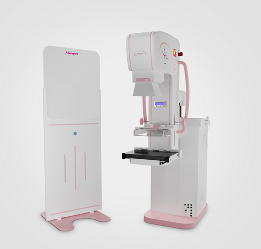
What types of scanners are used for Mammography?
- Digital Mammography (FFDM): Uses electronic detectors to capture clear breast images instantly on a computer.
- 3D Mammography (Tomosynthesis): Takes multiple images from different angles to create detailed 3D views, ideal for dense breasts.
- Analog Mammography: Older, film-based system (now mostly replaced).
- Contrast-Enhanced Mammography: Uses contrast dye for highlighting suspicious areas.
What Happens Before a Mammography?
Before a mammography, you’ll need to prepare to ensure clear and accurate images.
Scheduling
Try to book your mammogram a week after your menstrual period, when your breasts are less tender.
Inform your doctor or technologist if you are pregnant, breastfeeding, or have breast implants.
Clothing & Accessories:
Wear a two-piece outfit so you only need to remove your top.
Avoid wearing necklaces or jewelry around your chest or neck area.
Avoid Creams or Deodorants:
Do not apply deodorant, lotion, perfume, or powder under your arms or on your breasts on the day of the test — these can appear as spots on the X-ray.
Previous Records:
Bring any previous mammogram images or reports for comparison — this helps the radiologist identify changes over time.
Relax & Ask Questions:
The technologist will explain the procedure and make sure you’re comfortable before starting.
What Happens During a Mammography?
During a Mammography, your breasts are examined by X-ray using a special machine to capture detailed images of the breast tissue. The process is quick, safe, and usually takes about 10–15 minutes.
- Preparation:
- You’ll be asked to remove clothing from the waist up and wear a gown. Any jewelry or deodorant should be removed as it can interfere with the images.
Positioning:
The radiologic technologist will gently position one breast at a time on a flat plate of the mammography machine.
Compression:
. Another plate will lower to lightly compress the breast. This spreads out the tissue, reduces motion, and helps get clearer images with less radiation. You may feel mild pressure or discomfort, but it lasts only a few seconds
Image Capture:
X-ray images are taken from different angles. The technologist may ask you to hold your breath briefly during each image to avoid blurring.
Completion:
- The process is repeated for the other breast. Once done, you can get dressed immediately. The images are then reviewed by a radiologist, who prepares a report for your doctor.
How long does a Mammography take?
A mammography is a quick and simple test that usually takes about 10 to 15 minutes in total.
- The actual imaging time — when your breasts are positioned and X-rayed — takes only a few seconds per image.
- Most of the time is spent on positioning and preparation to make sure the images are clear and accurate.
- You may need to wait a few minutes afterward while the technologist checks that all images are properly captured.
What Happens After a Mammography?
Immediate Care:
You can get dressed right after the test.
Some women may feel mild tenderness in the breasts for a few hours due to the compression, but it usually goes away quickly.
Image Review:
The mammogram images are reviewed by a Radiologist, a doctor who specializes in interpreting breast X-rays.
The radiologist looks for any abnormalities, such as lumps, calcifications, or tissue changes.
Report Preparation:
The radiologist prepares a detailed report and sends it to your referring doctor or healthcare provider.
Results Discussion:
Your doctor will explain the results to you.
If anything unusual is found, you may be asked to return for additional tests, such as ultrasound, MRI, or biopsy, to confirm the findings.
What are the benefits of a Mammography ?
Mammography is one of the most effective tools for maintaining breast health and detecting problems early.
- Early Detection of Breast Cancer
- Mammography can detect small tumors or changes in the breast before they can be felt by hand.
- Early detection greatly improves treatment success and increases survival rates.
- Accurate and Detailed Imaging
- It provides clear, detailed pictures of breast tissue, helping doctors identify even tiny calcifications or abnormalities.
- Accurate and Detailed Imaging
- Modern digital and 3D scanners give high-quality images with minimal radiation.
- Quick and Non-Invasive
- The test takes only about 10–15 minutes.
- Quick and Non-Invasive
- No injections or recovery time are needed — you can return to your normal activities right after.
- Helps Differentiate Between Benign and Cancerous Changes
- Mammography can help doctors tell whether a lump is non-cancerous (benign) or needs further testing.
- Peace of Mind
- Regular screening offers reassurance and helps detect changes early, long before symptoms appear.
- Helps Differentiate Between Benign and Cancerous Changes
-
What are the risks of a Mammography ?
procedures, it has a few minor risks and limitations.
- Low Radiation Exposure Mammography uses a very small amount of X-ray radiation.
- The risk from this exposure is extremely low, and the benefits of early cancer detection far outweigh the risk.
- Temporary Discomfort The breast compression during the test may cause mild pain or discomfort, but it lasts only a few seconds.
- Some women may experience temporary tenderness afterward.
- False Positives Sometimes, a mammogram may show an abnormal result even when no cancer is present.
- This can lead to unnecessary worry or extra tests such as ultrasound or biopsy — though these are done only to be sure. Risks of a Mammography Mammography is a very safe test, but like all medical
- False Negatives In rare cases, especially with dense breast tissue, mammography may miss a small cancer.
- That’s why some women may also be advised to have breast ultrasound or MRI for additional screening.
- Anxiety and Stress Waiting for results or being called back for more images can cause temporary emotional stress, even if the findings turn out to be normal.
Care at North City Diagnostic
If you have concerns about your Breast Screening , you need a team of experts you can trust. Our Physicians and Radiologists at North City can help you with Mammography Of Breast