
Overview of MR ANGIO OF BRAIN
What is a MR Angio of brain ?
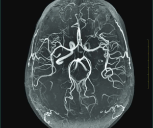
Magnetic resonance angiography of the brain is a non-invasive, painless diagnostic imaging procedure using radio waves and a strong magnetic field to create detailed images of the blood vessels in your brain.
A Brain MRI scan creates a picture of the brain bones (cranium), veins/arteries (grey/white matter), cranial nerves and surrounding soft tissues. The images captured during the MRI scan can be stored on an electronic device which is further printed on a film. The MRI scan is radiation less test which uses strong magnetic and radio waves to create detailed and clear 3D multiple images. The whole MRI scan procedure takes about 40-60 minutes.
What does an MR Angio of Brain diagnose?
An MR Angio (MRA) of the brain is a non-invasive imaging scan that primarily diagnoses issues related to the brain’s blood vessels and circulation.
Common diagnosis:
- Aneurysms: Weak, balloon-like bulges in blood vessels that could burst.
- Narrowed or blocked arteries: Blood vessels that don’t let enough blood flow to the brain.
- Stroke or mini-stroke (TIA): When blood flow to part of the brain is reduced or cut off.
- Abnormal blood vessel tangles (malformations): Unusual connections or clusters of blood vessels.
- Blood clots in brain veins: Blockages that stop blood from draining properly.
- Follow-up after treatment: Checking if surgery, stents, or coils are working well.
Indications:
- To check for bulging blood vessels (aneurysms) that could rupture.
- To see if any brain arteries are narrowed or blocked, which might reduce blood flow.
- To look for causes of a stroke or mini-stroke (TIA).
- To find out if there are any abnormal tangles of blood vessels.
- To check for blood clots in the brain’s veins.
- To monitor treatment results after surgery or stent/coil placement.
- To investigate unexplained symptoms such as severe headaches, dizziness, seizures, or vision problems.
- To screen people with a strong family history of brain aneurysms or vascular problems.
TEST DETAILS:
Who performs an MR Angio of Brain?
MRI technologist is responsible for performing the scan, operating the MRI machine, positioning the patient, and ensuring image quality.
Radiologist: A physician specialized in medical imaging who reviews and interprets the MRA images, provides the diagnosis, and issues the final report.
Nursing/support staff: May assist with patient preparation, IV contrast administration (if needed), and monitoring during the procedure.
What types of MRI scanners are used for MR Angio of Brain ?
Brain MRA is done using standard 1.5T or high-power 3T MRI scanners, sometimes with special methods like Time-of-Flight (no injection) or Contrast MRA (with dye), and open MRI may be used for patients uncomfortable in closed spaces though with less detail.
What happens before an MR Angio of Brain ?
Before a brain MR Angiography, the patient is asked about any medical history such as kidney problems, allergies, or implanted devices like pacemakers, metal clips, or stents, then they change into a hospital gown, remove jewelry or metal objects, and if contrast dye is needed an IV line is placed in the arm for injection during the scan.
What happens during MR Angio of Brain?
Magnetic resonance angiography may be done on an outpatient basis or during a hospital stay. Generally, magnetic resonance angiography follows this process:
- You will remove any clothing, jewellery, or other objects that may interfere with the scan and put on a gown.
- If you need a contrast dye to make blood vessels easier to see, this will be given through an IV.
- You will be positioned on an exam table directly outside the MRI scanner.
- The table will slide into position, placing you inside the MRI scanner.
- You will need to lie still during the scanning process. Any movements can blur the images and cause the results to be less accurate.
- The full scan may take half an hour to one hour or longer. This will depend on the type and number of blood vessels that your healthcare provider wishes to examine.
The scan typically causes no side effects or complications. If it is done on an outpatient basis, you are generally free to leave after the magnetic resonance angiography. Your healthcare provider will likely schedule a follow-up appointment to review the results of the test.
How long does an MR Angio in Brain take?
An MR Angiography of the brain usually takes about 30 to 60 minutes, depending on whether contrast dye is used and how many images are needed.
What happens after an MR Angio of Brain?
Your healthcare provider will examine the images from the magnetic resonance angiography. If no blockages or irregularities are found, you have what’s called a normal test result. An abnormal result means that the healthcare provider noted an abnormality in one or more of the blood vessels in your body. This may suggest that you have hardening of the arteries, known as atherosclerosis, or another circulatory problem. Your healthcare provider will likely suggest additional tests or treatments based on the specific problem that is discovered.
What are the benefits of an MR Angio of Brain ?
Brain MR Angiography is a safe, non-invasive scan that detects aneurysms, narrowed or blocked arteries, vascular malformations, and helps monitor treatment without using radiation.
What are the risks of an MR Angio of Brain ?
Brain MR Angiography is generally safe, but risks include allergic reaction to contrast dye (if used), kidney problems in patients with severe kidney disease, discomfort or claustrophobia inside the MRI machine, and rare issues with implanted metal devices that are not MRI-compatible.
A note from North City Diagnostic
The uncertainty of not knowing what’s happening with your body can make you uneasy. An MR Angio Of Brain can provide answers. his relatively low-risk contrast MR Angiography of the brain helps your healthcare provider detect problems like aneurysms, narrowed or blocked arteries, strokes, abnormal blood vessel tangles (vascular malformations), and blood clots, allowing for early diagnosis and treatment planning. Different types of MRI scanners are available, depending on your needs and preferences. Your provider will discuss next steps with you based on your test findings.
Care at North City Diagnostic
If you have concerns about your brain’s blood vessels or circulation, you need a team of experts you can trust. Our Physicians and Radiologists at North City can help with Brain MR Angiography.